Link t full russian text
Download PDF format
Russian Journal of Parasitology, 2017, V. 40, Iss.2
DOI:
Received 09.02.2017
Accepted 12.04.2017
CASE REPORT: CLINICAL SINGS LESIONS OF BLACK BAG DISEASE OF SARCOTACES SPР. (COPEPODA, PHILICHTHYIDAE) INFESTATION CROPPER FISH EPINEPHELUS TAUVINA IN NORTHWEST ARABIAN GULF
Essa T. Mohamed
Department of Marine invertebrate and marine Vertebrate, Marine Science Center, University of Basra, Iraq.
Abstract
Sarcotaces sp. (copepods), which infest and some the clinical sings lesions in cropper fish Epinephelus tauvina were register in Northwest Arabian Gulf. In March and June 2016, a total of 8 fish freshly collected from Al-Fao market South of Al-Basra City, northwest Arabian Gulf, Iraq. One of fish only infested with Sarcotaces sp. infested with Black Bag Disease with prevalence percentage 12.5 % Fish infected with Sarcotaces sp. showed no any clinical abnormalities and appeared to be completely normal. A new host and new geographical distribution for this copepod parasite in the fish of the Iraqi marine waters of the Arabian Gulf
Key words: Sarcotaces sp., Copepod, Marine Fish, Arabian Gulf, Epinephelus, Iraq.
Introduction
The family Philichthyidae Vogt, 1877 (Copepoda, Poecilostomatoida) comprises copepods that live in the pores of lateral lines and mucous canals of the mandibular and/or preopercular bones and cephalic canal system of fish hosts, At these sites, the copepods larvae penetrate their hosts and the females remain at the site of penetration for the rest of their lives. Males may enter the same site or go out when necessary, and are rarely found in the canal with the females. While in the host, the female undergoes a metamorphosis; characterized by the development of processes on some of the body segments, the thoracic legs in particular becoming diminutive (Boxshall & Mont, 1997; Boxshall & Halsey, 2004).The crustaceans can be harmful by its presence and the number of the parasites which is recognizable by black fluid of the parasite Eissa, I.A.M., (2002). The copepod Sarcotaces are usually found in galls, not visible externally (Woo, P.T.K., 1995).The aim of this study was the detection of the prevalence of Sarcotaces sp. in cropper fish form Northwest Arabian Gulf of Iraq.
Materials and methods
Parasitic copepod was carried out while the fish were fresh. Skin, fins, abdominal cavity, dorsal and lateral musculature was investigated macroscopically using magnifying lens and also with the help of a Stereomicroscope under 6 X magnification for the presence of copepods cysts of Epinephelus fishes. Fishes were collected from Al-Fao market South of Al-Basra City, northwest Arabian Gulf, Iraq. During March and June 2016. fishes were transported to the laboratory of Marine Science Center. The abdominal cavity of each fish was open and the intestine was separated from the other visceral organs and placed in a Petri-dish containing physiological saline. For each host specimen, the spiral intestine was removed, opened with a longitudinal incision, fixed in 10% formalin buffered with seawater, and transferred to 70% ethanol for storage.
Encysted Sarcotaces parasite were removed and put in Petri dish. The cyst was opened and preserved in 4% formalin for microscopic examination. Photographs were down using a Sony camera tube attached microscope.
Results and discussion
Sarcotaces sp., juvenile's fig 1
Class: Crustacea
Order: Poecilostomatoida
Family: Philichthyidae Vogt, 1877
Genus: Sarcotaces Olsson, 1872
Three cysts located within the flesh of on dead fish infected of one specimen's, directly beneath the skin in the same level of body surfaces wollen in the dorsal musculature or near the vent.
Cysts were pyriform in shape and gray in color (Fig.1). The juvenile was elongated, slender in shape, and flattened dorso-ventrally. Body segments are definable only by lateral notches. The head is small, nearly squarish and wide than long and separated from the thorax by clearly defined constriction. The first three thoracic segments are wider than the rest of the body.
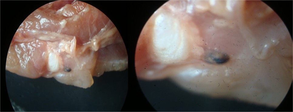
Figure 1: photograph showing Epinephelus tauvina musculature infected with Sarcotaces spр.
Black bag disease, a disease due to Sarcotaces (Copepoda, Philichthyidae) infection, has a worldwide distribution, from abyssal depths to tropical reefs (Amlacher, E., 1970; Boxshall G.A. and S.H. Halsey, 2004 ; Moser, M., L. Haldorson and J. Field, 1985).
The genus Sarcotaces has far been known to have four species. These are S. verrucosus (Olsson, 1872), parasitic on Acanthurus sp. from the West Indies, S. arcticus (Collett, 1874), parasitic on Molva abyssorum from Norway, S. pacijicus (Komai, 1924), parasitic on Antennarius sp., and S. komaii (Shiino, 1953), parasitic on Peristedion amiscus. The last species of Sarcotaces sp. (Yamaguti, 1963), taken from Semicossvphus reticulate belong to the species found in Japan. The Genus Sarcotaces is a bizarre copepods cause cysts formation was first reported by Heegaard, 1947 which measures several centimeters in length in the muscle under the skin.
Kuitunen-Ekbaum, (1949) and Roberts et al., (1988) who reported that, the cysts are exposed when fish are filleted and cut the cysts gall. The cyst of Sarcotaces is pyriform in shape, gray in color and release copious black fluid over the fillets. Hosts of these parasites include members of the families Moridae, Macrouridae, Scorpaenidae, Antennariidae, Triglidae, Acanthuridae and Serranidae (Izawa, 1974).
In this study, Sarcotaces sp. was recovered from cysts on the flank musculature, beneath the skin of Epinephelus tauvina (Family: Serranidae). A new host and geographical record for Sarcotaces is presented, being the first record of Sarcotaces sp. in the Iraqi marine fish species of the Arabian Gulf.
References
Amlacher, E.1958. Ein seltener und ein haufigerer parasitischer Krebs an Meeresfischen. Dtsch. Fisch. Ztg. 3: 75-77.
Amlacher, E., 1961. Taschenbuch der OCEAN, July 28-August 5, 1986. Canadian MS Fischkrankheiten fur Fischereibiologen, Tierarzte, Report of Fisheries and Aquatic Sciences, 1981. Fischzuchterund Aquarianer, VEB verlag Fischer, 29.Conzalez Raul, A. and Ruben D. Tanzola, 2000. On Jena, pp: 66-73. The presence of Sarcotaces verrucosus (Copepoda).
Boxshall G.A. And S.H.Halsey, 2004‘An Introduction to Copepod Diversity’ (The Ray Society: London).
Dollfus, R., 1928. Un hote noveau pour Sarcotaces verrucosus, Olsson, 1872 (Copepoda Parasite), Bulletin du Museum national d'Histoire Naturelle, Paris, 34: 341-345.
Eissa, I.A.M., (2002). Parasitic fish diseases in Egypt.El-Nahda El-Arabia Publisher, 32 Abd El-Khalek, Tharwat Street, Cairo, Egypt.
Kuitunen-Ekbaum, E., 1949. The occurrence of Sarcotaces in Canada. Journal of Fisheries Research Board of Canada, 7: 505-512.
Komai, T., 1924. Notes on Sarcotaces pacificus n. sp. with remarks on its systematic position. Mem. Coll. Sc. Kyoto Imp. Univ. S. B.1, 13(3): 273-83.
Roberts, R.J., 2012. Fish Pathology Fourth Ed., W.B.Saunders.An imprint of Harcourt Publishers.
Heegaard, P., 1947. Discussion of the genus Sarcotaces (Copepoda) with a description of the first known male of the genus. Kungl. fysiogr. Sallsk. Lund Forhandl. 17: 122-129.
Kabata, Z., 1970. Crustacea as enemies of fishes. In salmon de mar Pseudopercis semifasciata (Cuvier, Diseases of fishes. Edited by S. F. Snieszko and H. R. 1829) (Pinguipedidae) en el Golfo San Matias, Axelrod. T. F. H. Publication. New Jersey, pp.: 3-171. Patagonia, Argentina, Tesis Doctoral, Universidad.
Moser, M., 1977. Sarcotaces sp. (Copepoda) on the in the Southwest Atlantic Acta Parasitological, head of Physiculus rastrelliger frame Salvador.45 (4): 345-349.Canadian Journal of' Zoology, 55: 258-260.
Izawa, K., 1974. Sarcotaces, a genus of parasitic copepod parasites of deep-sea fishes. Journal of copepod (Cyclopoida:Philichthyidae), found on Marine Systems, 15: 215-223.
Roberts, R.J., D.J Macintosh, K.Tonguthai National Del Sur.S. Boonyaratpalin, N. Tayaputch, M.J. Phillips and 28. Stanley, R.1988.Cruise details and biologica.
S.D. Millar, 1986. Field and Laboratory Investigations observations from rockfish charters to the west coast into Ulcerative FishDiseases in the Asia-Pacific of Vancouver Island aboard the F/V HOWE BAY. Region. Bangkok: F.A.O. July 24-31 and August 24-29, 1985 and F/V ARCTIC
Eissa, I.A.M., 2002. Parasitic fish diseases in Egypt. El-Nahda El-Arabia Publisher, 32 Abd El-Khalek, Tharwat Street, Cairo, Egypt.
Woo, P.T.K., 1995. Fish diseases and disorders. CAB, Int. Wallingford, Oxon, UK.
© 2017 The Author(s). Published by All-Russian Scientific Research Institute of Fundamental and Applied Parasitology of Animals and Plants named after K.I. Skryabin. This is an open access article under the Agreement of 02.07.2014 (Russian Science Citation Index (RSCI) http://elibrary.ru/projects/citation/cit_index.asp) and the Agreement of 12.06.2014 (CA-BI.org/Human Sciences section: http://www.cabi.org/Uploads/CABI/publishing/fulltext-products/cabi-fulltext-material-from-journals-b...)